Proliferation and growth assay
Adherent cell culture proliferation assay in a serum comparative study with time-lapse imaging
Introduction
The term “cell growth” is used in the contexts of biological cell development, cell division (reproduction) and cell proliferation. Cell proliferation is an important indicator for assessing cellular activity, metabolism, physiology, pathology, normal tissue development, regeneration and renewal. There are several types of cell proliferation assays based on DNA synthesis (e.g. 3H thymidine incorporation, BrdU), on metabolic activity (LDH) or on ATP concentration (luciferase bioluminescence). Stem cells and cancer cells are very important in cell proliferation assays. Imaging allows to visualize cell proliferation and growth. This is particularly the case of IPRASENSE products.
The CYONOTE records cell growth with time-lapse imaging. Cells are automatically counted without labelling. Furthermore, with the CYTONOTE, it is no longer necessary to extract the cells at regular intervals to perform the counts. The HORUS software is creating a growth curve to monitor the growth of cells without extracting the medium.
Material and me
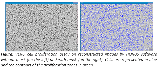
The CYTONOTE is able to perform measurements inside the incubator and it recognizes cells without any labelling. HORUS software automatically calculates cell number, cell saturation, cell area, cell concentration, cell morphology …
For the experiment, VERO cell line is cultivated in six-well plates in an incubator at 37 ° C with 95% humidity. Image acquisition was performed with the CYTONOTE 6W lens-free video microscope. As you can see on this picture, cell confluence can be observed and measured
Results
The CYTONOTE 6W is designed to monitor the 6 sensors simultaneously or distinctly for 6 parallel or independent cell cultures. The experiment was performed in duplicate with 3 different conditions by adding 3 different serums. The CYTONOTE carried out continuous measurements during 4 days from inoculation (day 0).
Between days 0 and 1, the HORUS software gives repeatable results, as shown with the overlapping curves for the same conditions. On day 1, three different serums were added to the cell culture. We see that the proliferation curve is different according to the serum because the speed of growth is different. The proliferation of the culture in contact with the serum A is faster than that with the serum B. That in contact with the serum C is faster but more regular than the other two cultures. Furthermore, the curves are always overlapped for the same conditions.
The CYTONOTE 6W allows us to compare proliferation of VERO cell line that had been in contact with 3 different serums. Thus, serum has an impact on cell proliferation.
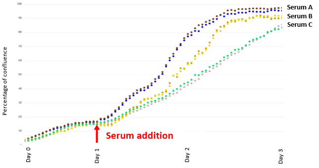
Figure 1 : The NORMA XS versus the Vi-Cell XR Evaluation on perfusion and fed-batch processes (data courtesy of GSK)
Conclusion
Performing real-time monitoring of cell proliferation instead of endpoint measurement provided significant advantages. The CYTONOTE allows to visualize and quantify cell proliferation using time-lapse imaging. Analysis of growth curves is automatic with the HORUS software and doubling times can be extrapolated. The CYTONOTE 6W is an easy tool to perform monitor cell growth.