Live Cell Imaging inside the Incubator
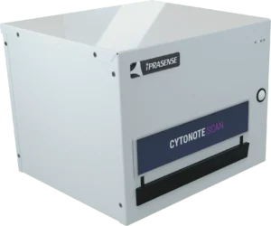
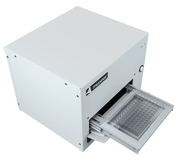
CYTONOTE SCAN
Time-lapse imaging of cell culture and multiwell plate analysis from inside the incubator
The CYTONOTE SCAN is designed for parallel cultures monitoring in multiwell plate
For research use only (RUO). Not for use in diagnostic procedures.
Label Free & High Contrast

Always in Focus

Settings Free
Huge Field of View
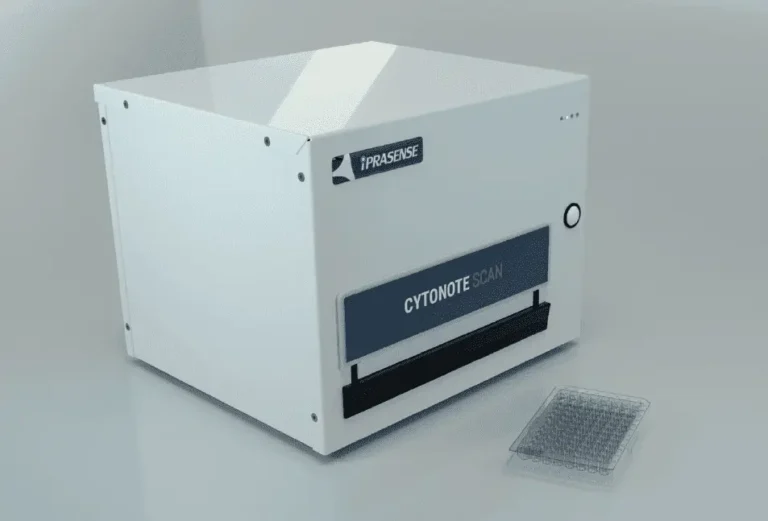
Live Cell Imaging System for Multiwell Plates from inside your Incubator
The CYTONOTE SCAN, based on our patented lens-less imaging technology, is the simplest live cell-imaging system designed for recording cell movies and analyzing a variety of cell culture in parallel from inside the incubator. The innovative and patented “Lens-less imaging” technology pushes the boundaries of microscopy with its super wide field of view and its capability to capture and analyze precisely several thousands of cells without any focus and brightness settings. This technology simplifies the Live Cell Imaging technique and transforms the complex and expensive microscope method into a cost-effective solution.
Features of our Live Cell Imaging System
- High contrast
- Always in focus
- Settings free
- Huge field of view
- Cell confluence determination
- Cell based assay: proliferation, migration, tacking, wound, healing…
- Clone selection
- 6, 12, 24, 96, 384 well-plate cell cultures + Petri Dishes
Applications of our Live Cell Imaging System
- Real-Time Imaging of your cell culture
- Designed for 6, 12, 24, 96, 384 well-plate cell cultures
- Time-Lapses & Movies
- Confluence analysis
- Growth curves
- Migration test
- Cell morphology
- Clone selection
- Proliferation test
- Cell population
- Cell attachment / detachment
- Cell tracking
Accessories & Consumables of our Live Cell Imaging System
- HORUS Software
- 21 CFR Part 11
- Scratch Assay Analysis module for HORUS
- Cell Tracking Analysis module for HORUS
- Cell Tube Formation Analysis module for HORUS
- CYTONOTE Cable (2 meters)
- Laptop Computer
- IQ/OQ
How to use
our Multiwell Live Cell Imaging System ?
The CYTONOTE SCAN is the simplest live cell-imaging system for multiwell plates designed for recording cell movies and analyzing a variety of cell culture from the incubator. The CYTONOTE SCAN is the perfect instrument for the monitoring of your Petri Dishes and 6, 12, 24, 96 and 384-well plate cell culture. The acquisition time between two measurements is 20 minutes by default, but you can change this time if you wish. By performing acquisitions over a given period of time you will be able to obtain time-lapses and movies of your cells.
As explained, no need to adjust focus, brightness or any other parameters, our algorithm will reconstruct cells wherever cells are located… from few micrometers to some millimeters! This characteristic makes the CYTONOTE SCAN an extremely robust device not fearing temperature variation, cells descent inside matrigel, or any other parameter which can affect traditional focus adjustment.
What are the results
of our Multiwell Live Cell Imaging System ?
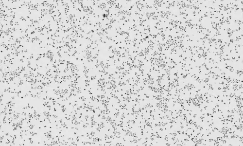
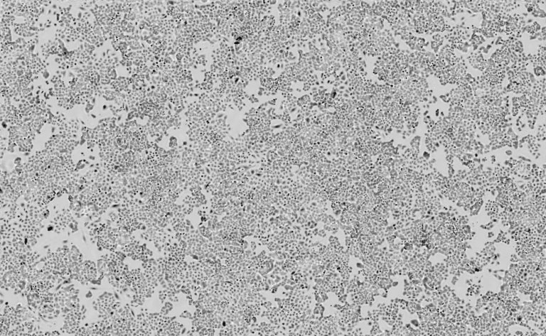
The CYTONOTE SCAN, with its wide field of view, converts all the data obtained into a single, representative image of the cell culture.
CYTONOTE SCAN images and results are analyzed using dedicated HORUS software.
HORUS is an application-oriented software package for determining confluence, cell size and cell tracking. Images are stored and can be viewed and zoomed at any time.
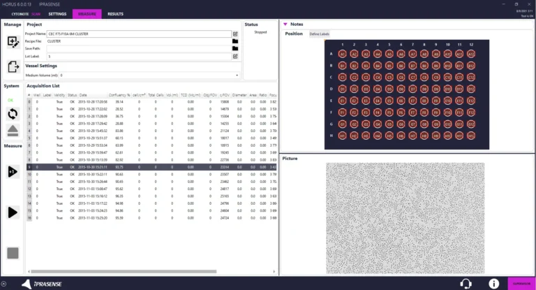
HORUS Software for our Live Cell Imaging System Included
Our best software for cell counting and cell analysis !
HORUS Software is included with the acquisition of our products and is regularly updated by our development team.
Technical Specifications
of our Live Cell Imaging System
Eucaryotic cells : Adherent monolayer, Suspension cell at bottom of culture ware or in micro-slides, 3D spheroids
Liquid or Semi-solid (collagen)
Standard 6, 12, 24, 48, 96, 384 mulwell plate, petri dishes
1 micron
30 mm² (6,5 mm x 6,5 mm)
0 to 5 mm
96 well plate in 15 min
LED
CMOS 10 Mpix
- .PNG
- .JPEG
- .BITMAP
- .TIFF
- Stainless steel
29,5 * 26,5 * 29,5 cm
12 kg
USB + 24 V DC (110 – 240 V AC)
- 21 CFR part 11
- IQ/OQ
FAQ
A Live Cell Imaging System is a cutting-edge technology that allows researchers to observe and analyze living cells in real-time within their natural environment, such as inside an incubator.
The CYTONOTE system offers high contrast, always-in-focus images, settings-free operation, a huge field of view, and robust performance.
With its patented lens-less imaging technology, the CYTONOTE eliminates the need for manual adjustments like focus and brightness settings. It provides hassle-free monitoring of up to 6 cell cultures in parallel.
The CYTONOTE system enables real-time imaging of cell cultures, time-lapses, confluence analysis, growth curves, migration and proliferation tests, cell morphology studies, cell tracking, and more.
Simply place your cell culture container, such as a Petri dish or T-flask, onto the CYTONOTE 1W. The system operates with a default acquisition time of 20 minutes between measurements, which can be adjusted as needed.
The CYTONOTE provides ultra-wide field-of-view imaging, with data analyzed using the dedicated HORUS software. Results include quantitative confluence determination, cell size analysis, cell tracking, and more.
By this product technology, there is a wide field of view, no manual adjustment step and the results are obtained in seconds.
The CYTONOTE supports various cell types and media, offers a resolution of 1 micron, a field of view of 30 mm², and operates with a CMOS 10 Mpix sensor. It’s compact, lightweight, and compatible with standard culture vessels.
Yes, accessories include cables, software modules for analysis (such as Scratch Assay and Cell Tracking), and compliance options for pharmaceutical industries (like 21 CFR part 11 and IQ/OQ).
Yes, all our products are 21 CFR.