Cell migration assay
Automated continuous monitoring of cell migration (Scratch assay) for senescence study
Introduction
Cell migration is a central process in the development and maintenance of multicellular organisms. Tissue formation during embryonic development, wound healing and immune responses all require the orchestrated movement of cells in particular directions to specific locations. Cells often migrate in response to specific external signals, including chemical signals and mechanical signals. Errors during this process have serious consequences, including intellectual disability, vascular disease, tumor formation and metastasis. An understanding of the mechanism by which cells migrate may lead to the development of novel therapeutic strategies for controlling, for example, invasive tumor cells. Cell migration and invasion play a role in many normal and pathological processes including immune responses, embryonic development, angiogenesis, regeneration, tumor metastasis and wound healing.
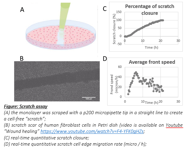
In vitro experiments, Scratch assay is the primary method for assessing cell migration and cell invasion. in this note of applications, we will describe this test through measurements with the CYTONOTE.
Cell migration assay
Cell culture
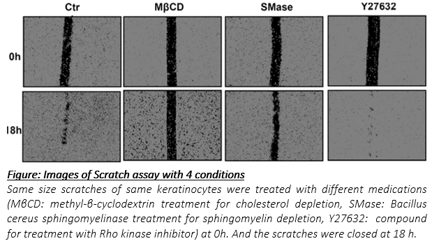
For the experiment, human epidermal keratinocytes were cultured in Petri dish of 35 mm² until confluence in an incubator at 37 ° C with 95% humidity. Then, the monolayer was scraped in a straight line to create a cell-free “scratch”. Keratinocytes in the scratch assay were first treated with mitomycin C (10 μg/ml) to inhibit cell proliferation while preserving viability and ability to migrate.
The CYTONOTE is able to perform measurements inside the incubator and it recognizes cells without any labelling. The HORUS software automatically calculates cell number, cell saturation, cell area, cell concentration, cell morphology … Unlike a conventional microscope, the absence of focus in the CYTONOTE allows an extremely wide field of view.
Results
While cholesterol depletion by MβCD (7.5 mM) abolishes keratinocyte migration, degradation of sphingomyelin in keratinocyte plasma membrane by incubation of cells with SMase (5 mU/ml) clearly slows down the process. Conversely, Y27632 which preserves the organization of sphingomyelin rich submicrometric domains during senescence is well known to simultaneously enhance cell migration, suggesting again possible links between sphingomyelin organization in keratinocyte membranes and their propensity to migrate. We observe the ability of keratinocytes to migrate decreases in senescent cells as well as in cells treated to deplete cholesterol or sphingomyelin in membranes.